Prostate cancer detection, diagnosis and treatment is an ever evolving field. Gone are the days of solely relying on PSA tests, DRE’s (Digital Rectal Exam) and blind TRUS biopsies. We believe in the following course of action as the best way for a man to determine the extent of his disease and make an informed decision on the best course of treatment.
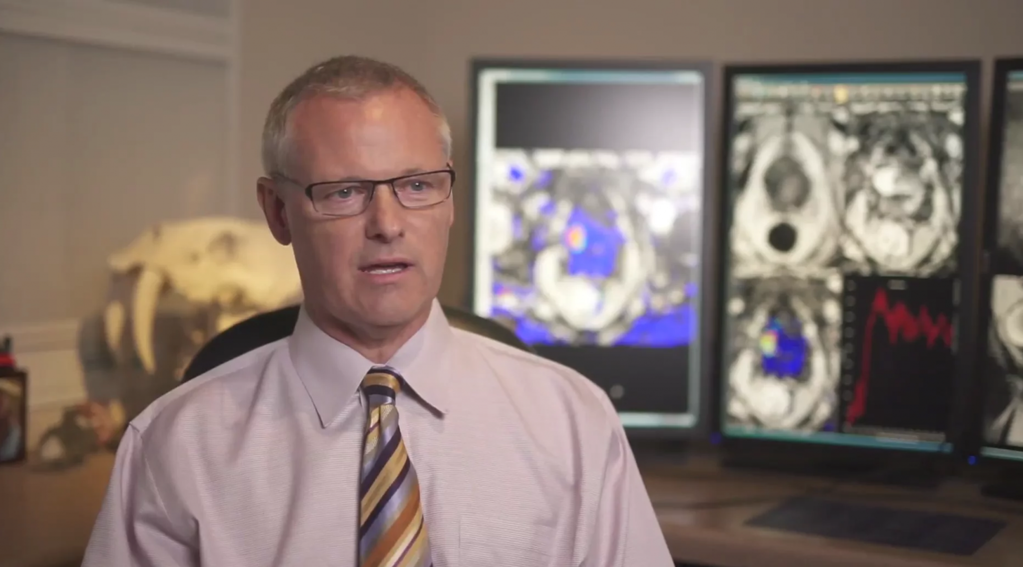
When a man is suspected of having prostate cancer, as a result of a rise in PSA, positive DRE or urinary symptoms, we at Desert Medical Imaging (DMI ) believe in imaging before biopsy. This means getting a multi-parametric MRI (mpMRI) scan of the prostate to identify any areas that might be suspicious for clinically significant cancer. At DMI we use state of the art MRI scanning technology to image the prostate which includes the use of a phased array pelvic coil. We do not use an endo-rectal coil.
If a suspicious area is found on the mpMRI scan, we can perform an MRI-guided biopsy that is targeted to the suspicious area. A targeted MRI-guided biopsy provides greater accuracy in diagnosis with fewer needle cores being taken, resulting in less pain and a lower risk of infection. An MRI guided biopsy takes place while the patient is “in-bore” inside the MRI machine. The biopsy needle is guided to the suspicious area and provides a record of exactly where the core or sample was taken. An mpMRI scan and MRI guided biopsy are invaluable tools for treatment planning, no matter which course of treatment you choose.
In 2010 the team of Bernadette Greenwood, BSc, PG Cert., RT(R)(MR) and Roger J. McNichols, PhD developed a unique approach using the Visualase® Thermal Therapy System and the Invivo DynaCAD/DynaTRIM prostate biopsy system to perform Laser Interstitial Thermal Therapy (LITT) or Focal Laser Ablation (FLA) and began conducting an IRB-approved Clinical Trial, NCT 02243033. The first case in the world was performed by Dr. John Feller, Dr. McNichols and Ms. Greenwood in May, 2010 as part of a Phase I Clinical Trial. The procedure involves placing the patient inside the MRI scanner and displaying MRI images and thermal maps on a computer screen. A thin laser fiber is guided to the tumor and laser energy is applied to heat, coagulate and kill the cancer cells. The temperature and the extent of the ablation zone are constantly monitored with MR imaging. The result is the elimination of MRI-visible cancer with very low risk of side effects. If it is determined through an mpMRI scan and MRI guided biopsy that you might be a good candidate for the procedure, this might be a treatment option you may wish to consider.
The Prostate Program at Desert Medical Imaging has further expanded to include tissue-based genomics, PET/CT Clinical Trials and blood tests for circulating tumor cells (CTCs).
The research team at DMI is dedicated to providing the utmost care in diagnosing and treating prostate cancer. The team includes:
In memoriam and for his contributions to this pioneering work, we recognize the efforts and talent of Dr. Stuart T. May, who passed away on January 22, 2017.
When my first Urologist suggested I use his procedure to remove my prostate, I decided to look for other alternatives. In my search, I came across this ablation procedure and thought that it would work best for me. I contacted DMI and set up and appointment and after initial testing and imaging, decided to go with it. I’m so glad I did. It’s been 10 months since my procedure and PSA is down to 1.4 now, and the images show a clean and healthy prostate gland. Everyone on the staff and those who worked directly with have been an absolute joy to work with.
Since April 1 when Dr. May located the small tumor in the left anterior lobe, a second tumor had started in the right anterior lobe. Because of early detection by the MRI biopsy, the cancer was contained and removed before it had a chance to spread. Several of my physician friends have confirmed that it was very rare to detect such an aggressive cancer in the anterior lobe this early and the routine ultrasound biopsy would not have caught it. Months of waitful watching could have been deadly.